Radiology
● Radiographs
● Ultrasound
● Mammograms – Tomosynthesis
● Bone Density Measurements
● CT Scans
● Magnetic Tomography
● Specialized heart imaging tests (Heart MRI – CT Coronary Imaging).
Based on valid and timely diagnosis and in collaboration with the doctors of other specialties, the most suitable imaging technique is selected in combination with appropriate protocols, observing all radiation protection rules, in accordance with international guidelines and the guidelines of the Hellenic Atomic Energy Commission.
Human resources consist of:
● Radiodiagnostics – Interventional Radiologists
● Radiophysicists
● Radiology/radiology technologists
● Medical imaging machine operators
● Nurses
● Secretaries
Modern equipment
Already in 2008 and keeping up with the evolution of technology, the Radiodiagnostics department has a digital diagnostic image archiving and communication system (PACS) for all examinees (electronic examination file – DICOM images), which significantly improves the daily work flow and makes a decisive contribution in optimal functioning and creative collaboration with physicians of all specialties.
Also, as the main core of its equipment, St Luke’s has the following:
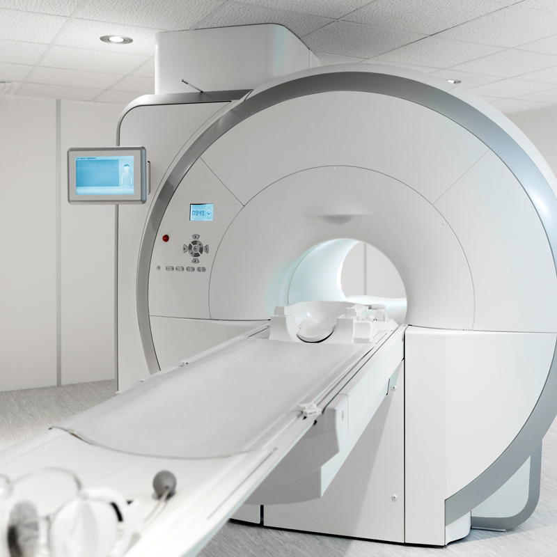
- State-of-the-art Magnetic Tomograph MAGNETOM Avanto fit 1.5 Tesla, Siemens
- Siemens Definition Dual Source 128 slices CT scanner
- 2 state-of-the-art Philips ultrasounds
- State-of-the-art Digital Mammogram – Siemens Tomosynthesis
- Bone mineral density scanner
- Angiographer
Examinations conducted:
Cardiovascular System Imaging:
- Cardiac CT- Coronary CT, CT Angiography before TAVI
- Magnetic Tomography of the Heart (in combination with the modern parametric sequences T1 mapping, T2 mapping, T2* mapping and specialized techniques for the estimation of myocardial strain parameters and the quantification of flows)
- Stress Magnetic Tomography of the Heart for the bloodless demonstration of myocardial ischemia, without radiation (in contrast to cardiac scintigraphy)
Nervous System Imaging:
Axial and Magnetic Tomography of the Brain, Axial and Magnetic Tomography of the Spine, Axial and Magnetic Angiography, MRI Perfusion, Spectroscopy, CSF (cerebrospinal fluid) Flow Measurement, Neuro-Navigation Protocol, fMRI, Epilepsy Protocol, Diffusion Tensor Tractography
Breast Imaging:
Breast Ultrasound, Digital Mammography-Tomosynthesis with 3D Imaging, Stereotactic breast biopsy and Ultrasound-guided biopsy
Pediatric Imaging:
All indicated radiological examinations, ultrasound examinations (abdominal ultrasound, inguinal ultrasound, testicular ultrasound, brain ultrasound, hip ultrasound, ultrasound-cystography, etc.), MRI or CT scans with personalized special pediatric protocols.
- Neck-Chest-Abdomen Imaging
- Imaging of the Genitourinary System
- Imaging of the Musculoskeletal System
- Oncological imaging
- Whole-Body MRI
- Axial Arteriovenous Angiography (e.g. Cerebral vessels, Carotid-Spinal Arteries, Thoracic and Abdominal Aorta, Pulmonary vessels, Renal Arteries, Upper and lower extremities, etc.)
- Arteriovenous Magnetic Angiography
- (Cerebral vessels, Carotid-Spinal Arteries, Thoracic and Abdominal Aorta, Renal Arteries, Upper and lower extremities, etc.)
- The entire spectrum of ultrasound screenings (thyroid, abdominal ultrasound, vascular ultrasound, soft tissue ultrasound, etc.)
- Guided Biopsies under CT or Ultrasound
- Axial Colonoscopy
The vision of the department
Our vision is to continue to offer high quality care for our patients by applying innovative techniques to the hospital‘s care system. Our diagnostic and therapeutic approaches always focus on the patient and their needs. This is achieved through the personal contact and respect with which we approach each patient and their individuality, understanding their concerns and contributing to their best possible guidance, aiming at the optimal therapeutic strategy.
Research and continuous training of the staff
In the Radiodiagnostic Department, the continuous training and education of the staff is one of the main priorities, as it allows us to look to the future and take into account all the current developments in the field of Radiology. Alongside the daily clinical practice and in collaboration with other scientific groups, we participate in research works by publishing the corresponding scientific articles, while our presence in Greek and international conferences is particularly active, but also noticeable.
Patient-centered collaboration
The Radiologists of St Luke’s work in close cooperation with many Doctors of various specialties (Cardiologists, Cardiac Surgeons, Pathologists, Urologists, Paediatricians, Pediatric Surgeons, Breast Surgeons, General Surgeons, Neurologists, Neurosurgeons, Otorhinolaryngologists, Oncologists, Gastroenterologists, Orthopedists, Ophthalmologists Pulmonologists, Thoracic surgeons, etc.), offering high-level services, ensuring the best possible care for patients.
The Radiology department of St Luke’s, with willingness, love and readiness, supports and promotes the treatment of emergencies, both inpatients and outpatients, in the following ways.
24 hours a day, there is a specialist Radiologist, who, supported by the complete and state-of-the-art technical equipment and the highly trained, experienced and cooperative technologists-operators of the radiological machines, safely and confidently offers the information that the treating doctor needs.
In cases of acute abdomen, if there is an indication after the abdominal x-ray, they approach the patient’s problem initially with an ultrasound:
In pediatric patients, to investigate acute appendicitis, intussusception, pyelonephritis, etc. Ingestion of a foreign body in children, initially a chest and abdominal x-ray. If there is an indication, a CT scan is performed immediately.
Also carrying out a CT scan in adults to investigate any ileus, find the cause of renal colic, investigate any visceral rupture, air leak.
Injury after a traffic accident or any other cause, to highlight any rupture of the intestines.
In cases of any C.E.C., exclusion of bleeding or fracture by computed tomography.
Similarly, in acute musculoskeletal injuries, depending on the case, perform a simple X-ray, CT or MRI.
In the acute setting of hemorrhagic or ischemic stroke, perform brain MRI. Likewise for first-onset epileptic seizures.
For any deterioration in the health of a patient in the hospital, it provides immediate information according to the examination requested by the attending physician.
In case of pulmonary embolism, even in small branches of pulmonary arteries, CT control is immediately performed with state-of-the-art 128-section equipment.


